Skip to content
University of Surrey, Guildford, Surrey, GU2 7XH, UK.

Mon to Fri: 8:30am – 5:30pm

Linkedin Youtube
Menu
  • Home
  • About us
    • Research & Development
      • Research Areas
      • Publications
    • Impact
    • Our People
    • Outreach & Training
    • Our History
  • Equipment & Facilities
    • Ion Implantation and Irradiation
    • Ion Beam Analysis
    • Secondary Ion Mass Spectrometry
    • Deterministic Implantation Backup
  • Getting Access
  • News & Events
  • Gallery
  • Contact
Feedback

Gallery

  • All
  • Staff
  • Ion Implantation​
  • Single Ion Implantation​
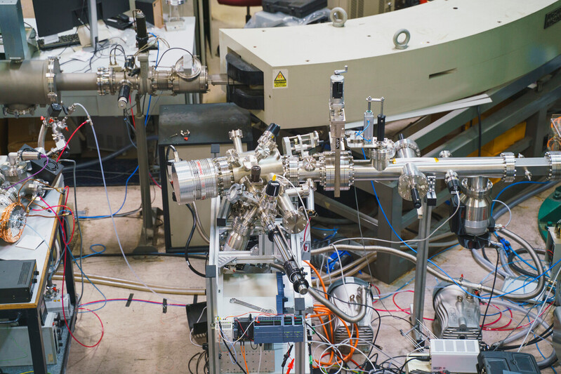
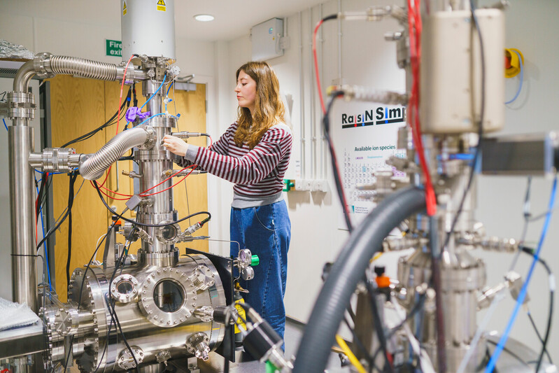
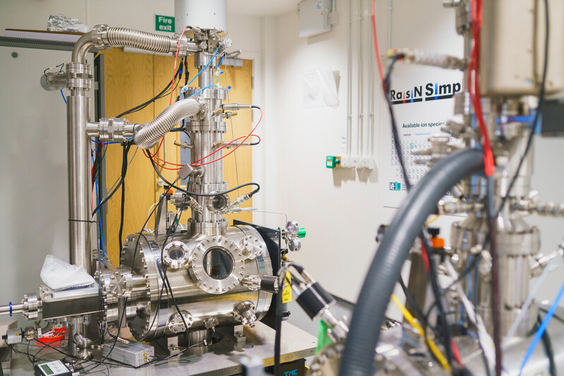
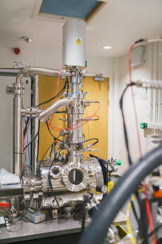
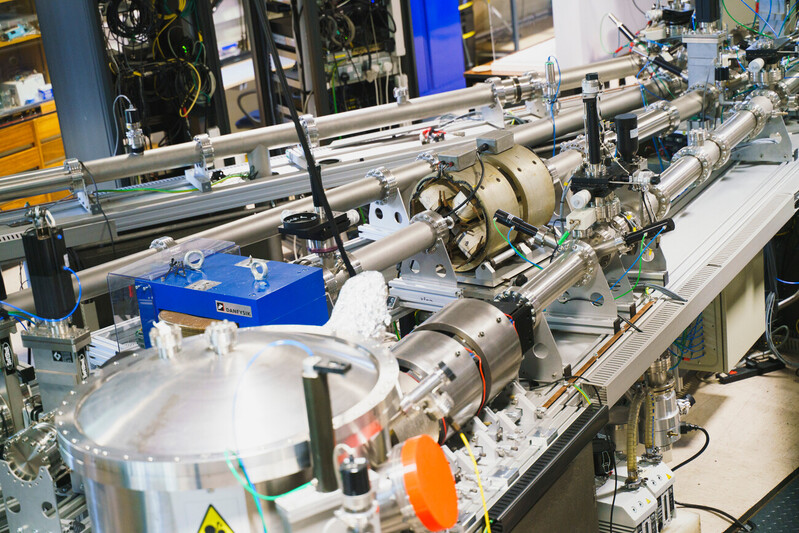
  • Ion Beam Analysis​

Mail

Luke.Antwis@surrey.ac.uk

Call Us

+44 (0)1483 689145

Address

Surrey Ion Beam Centre, University of Surrey,
Guildford, Surrey,
GU2 7XH, UK.

© 2025 ionBeam. All Rights Reserved, Site By Vidvath.

  • Terms & Privacy

6. Decision

If the Validator declines to validate, they are asked for a reason by the UKNIBC, which communicates this to the proposer. But if the proposal is validated, the project starts immediately.

5. FAF validation

The Validator (if required) is not a referee, although s/he is welcome (and encouraged) to make comments on proposal quality. Indeed, such comments will be helpful to rank proposals in the event that the UKNIBC is oversubscribed.

However, the UKNIBC funding is classed by EPSRC as already refereed, so the primary purpose of the Validator is purely as an independent confirmation that UKNIBC resource is being applied appropriately.

4. FAF creation

The FAF will contain all appropriate details, including the user’s name (and also the PI’s, if it is different). A Summary Statement of the Grant Holder shows how the proposed project is related to the EPSRC grant named. A Summary of UKNIBC Contribution to the Project includes the specifics of the work proposed at the Facility. This should be detailed enough to justify the resource requested. ​

3. Request refinement

The request is further refined following the review by the team. The Discussion will be documented (via email), and may be as detailed and extended as required.

2. Request review

The request is reviewed. Someone at the Local Facility (the Liaison Fellow, LF) is chosen to discuss the request with the user through the contact details shared during request submission.

1. Request initiation

Potential users can approach the facility without any introduction. Conversely, Facility staff may have encouraged their contacts.

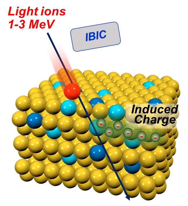
Ion Beam Induced Current (IBIC) : Measures the charge collection efficiency or current generated in an electronic or semiconductor device when irradiated with an ion beam. IBIC provides spatially resolved maps of electrical activity, carrier transport, and radiation damage, enabling the evaluation of detector performance and semiconductor device integrity.

  • Applicable to the analysis of devices and planar semiconductor or insulator wafers, which may contain grain boundaries, dislocations or other types of lattice defects
  • Can be run simultaneously with PIXE, RBS and IBIL

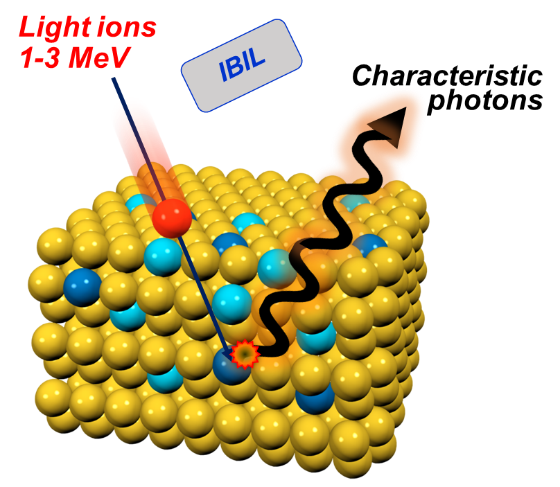
Ion Beam Induced Luminescence (IBIL) : Detects visible or near-visible photons emitted from the sample as a result of ion excitation. The luminescence spectrum reveals information about electronic structure, impurities, and defect states, making IBIL a valuable tool for studying semiconductors, minerals, and scintillating materials.

  • Light is emitted as a result of outer-shell electron transitions, giving information about molecular bonds in the material
  • Potential to probe up to ~20 μm below the surface

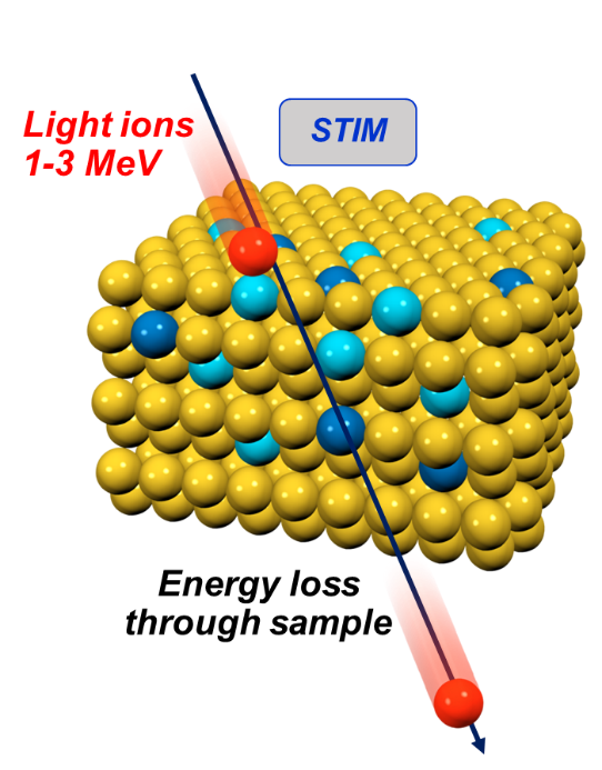
Scanning Transmission Ion Microscopy (STIM) : Uses a finely focused MeV ion beam scanned across a thin specimen. By measuring the energy loss or transmitted particle intensity, STIM provides quantitative two-dimensional or three-dimensional maps of areal density and thickness at sub-micrometer spatial resolution. It is particularly useful for biological samples, thin films, and microelectronic structures.

  • Technique allows measurement of energy loss through thin layers of material
  • Energy loss of primary beam increases with increasing atomic mass
  • Can be run simultaneously with PIXE and RBS

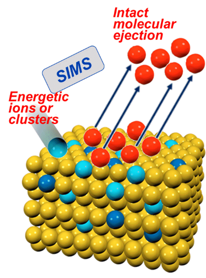
MeV Secondary Ion Mass Spectrometry (MeV-SIMS) : Combines the principles of SIMS with high-energy (MeV) primary ions. The electronic stopping processes at these energies produce significantly higher secondary ion yields with minimal fragmentation, allowing for molecular analysis while preserving depth resolution. MeV-SIMS bridges the gap between traditional SIMS and nuclear-based IBA, offering chemical and molecular information alongside elemental quantification.

  • MeV beams can be focused and scanned several centimetres into air.
  • Molecular mapping.
  • Highly desirable for large samples that cannot be placed in a vacuum chamber.

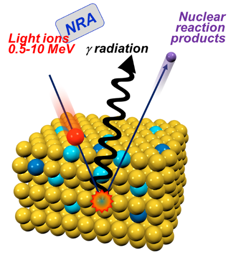
Nuclear Reaction Analysis (NRA) : Uses specific nuclear reactions to identify and quantify elements with high sensitivity.

  • The technique is particularly useful for identifying light elements that are difficult to “see” with PIXE
  • NRA is a nuclear to obtain concentration vs. depth distributions for certain target chemical elements in a solid thin film.
  • Resonant reactions can be used which occur at particularly sharp interaction energies. As the mono-energetic beam slows gradually as it enters the target, these resonant reactions occur only at determinable depths. Hence by varying the energy of the initial beam it is possible to depth profile using this technique.

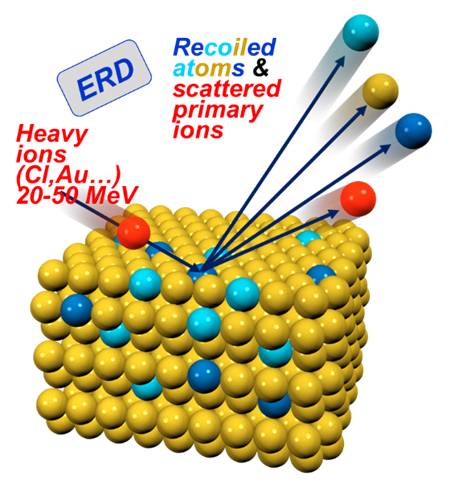
Elastic Recoil Detection Analysis (ERDA) : Detects atoms recoiling from the surface after ion impact, particularly useful for quantifying light elements such as hydrogen.

  • Depth profiling of light elements and isotopes (e.g. H and D)
  • Depth resolution of 10 nm (except for H and He)
  • Detection limits of 0.1 at% in volume, 5×10¹⁴ at/cm2 as thin film
  • Heavy ion ERD (10-100 MeV) and light ion ERD (~MeV)
  • Good mass resolution for lighter atoms
  • Multi-elemental stoichiometry in complex matrices or multilayer films (including polymers)

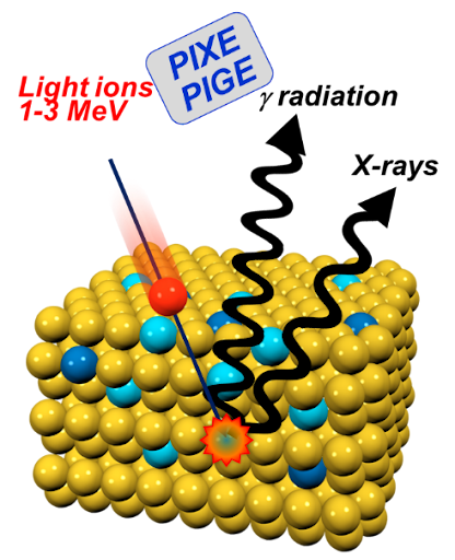
Particle-Induced X-ray Emission (PIXE) : Detects characteristic X-rays emitted when the incident ions ionize inner-shell electrons, allowing for multi-elemental analysis with high sensitivity.

  • PIXE can be used to detect elements in the periodic table from Na to U with sensitivity is at the ppm level for most elements.
  • PIXE analysis gives absolute quantification,  there is no need to compare to similar standards.
  • Accuracy of the technique is ~5%
  • Analysis can be carried out in air. This allows the non-destructive analysis of large objects.
  • Combination with RBS gives us complementary information that we can use to determine depth profiles of some major elements, and information about light elements.

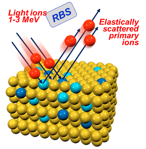
Rutherford Backscattering Spectrometry (RBS): Measures the energy of ions scattered elastically from nuclei, providing information on elemental composition and depth distribution.

    • RBS determines compositional information about a sample
    • Atomic concentration of major and minor elements with absolute accuracy (<1% at best)
    • Accurate determination of film thickness
    • Depth profiles with ~10nm resolution
    • Sensitivity ~at% (a few monolayers at the surface)
    • No sample preparation is required and not deliberately destructive
    • Can be carried out in air and simultaneously to PIXE, NRA and ERD

    UNIVERSITY OF SURREY ION BEAM CENTRE

    University of Surrey Feedback Form

    Our “Quality Objectives” include:

    • If we make a mistake, we will acknowledge it and correct it quickly.
    • We analyse customer feedback to ensure quality objectives are achieved.

    Please complete this form to help us improve. You may remain anonymous.

    FEEDBACK FORM

    How would you rate the following:

    The work was undertaken in a timely way
    The work was undertaken correctly
    The work was specified correctly
    The IBC staff were helpful
    The IBC collaboration added value
    This work is expected to lead to outputs